Ophthalmic Manifestations of Feline Herpes Virus Type-1
By Dr. Amanda Corr, VMD, DACVO | Ophthalmology
Feline Herpes Virus type-1 (FHV-1) is the most common cause of ophthalmic diseases affecting cats. FHV-1 is species specific and highly contagious. Over 90% of cats are seropositive for the virus. Most cats are exposed to the virus between 8 and 12 weeks of age and contact the virus via direct contact, aerosolization or fomites. The virus can be passed from the mother during gestation, or shortly after birth. Severe infection within the first two weeks of life, prior to eyelid opening, can cause neonatal ophthalmia which commonly leads to corneal rupture.
Primary FHV-1 infection can also occur in older kittens or adults. 80% of infected cats remain latently infected for life with virus sequestered in the trigeminal nerve ganglion. Almost half of those cats shed the virus for life.
Vaccination against FHV-1 with modified live (parenteral) or killed (oronasal) vaccines only provides limited and temporary immunity. Despite vaccination cats can be re-infected with the virus. However, recurrent disease is most commonly thought to be associated with recrudescence of latent virus.
Chronic or recurrent ocular disease associated with FHV-1 is extremely common. Flare-ups may be associated with stress such as illness/hospitalization or change in environment. Clinically, an obvious stressor is often not identified and onset of active disease appears to be random.
Mechanisms of FHV-1 induced disease
FHV-1 infects epithelial cells within the respiratory tract, conjunctiva and/or cornea. During primary infection or when the virus is reactivated following latency, the direct effect is cytolysis of the infected cells and necrosis of the affected tissues. Additionally, inflammatory disease can occur via an immune-mediated response to the infection.
Clinical manifestations of disease
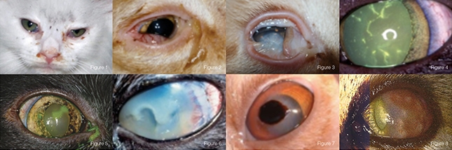
In kittens, primary FHV-1 infection leads to upper respiratory disease (viral rhinotracheaitis) as well as ocular disease. In addition to nasal and ocular discharge, these kittens may act sick with fever, generalized malaise, decreased appetite, sneezing and coughing (Figure 1). Purulent nasal and ocular discharge can be present with viral infection alone, however, secondary bacterial infection is common. Most commonly, primary infection is ‘self-limiting’ meaning the kitten’s immune system is able to clear the disease within 10-14 days. However, the course of the disease can be extremely variable. In adult cats or cats with recurrent infection, clinical signs are typically less severe and only involve the eyes.
Conjunctivitis is the most common disease caused by FHV-1 and is the most common feline ophthalmic disease. Most cats with conjunctivitis are blepharospastic with ocular discharge, conjunctival hyperemia and chemosis (Figure 2). Clinical signs can range from mild to severe and ocular discharge can range from serous/epiphora to mucoid to purulent. Cats with ulcerative conjunctivitis tend to be more painful. In young kittens, ulcerated conjunctiva can heal to itself or to corneal ulcers, forming symblepharon (Figure 3) which can lead to severe permanent visual deficits. Chronic conjunctivitis can also lead to scarring of the palpebral conjunctiva and entropion.
FHV-1 can infect corneal epithelial cells and cause painful corneal erosions or ulcers. Dendritic corneal ulcers are pathognomonic for Feline Herpes Virus 1 infection (Figure 4). Corneal erosions do not take up fluorescein stain but do stain positive with Rose Bengal. FHV-1 infection can also lead to larger, geographic corneal ulcers which are commonly surrounding by non-vital epithelium (Figure 5).
Superficial corneal ulcers secondary to FHV-1 can progress rapidly to stromal corneal ulcers (Figure 6), descemetoceles and corneal perforation. Chronic superficial corneal ulceration due to FHV-1 infection can cause sequestra, or scab-like lesions of necrotic corneal tissue (Figure 7). Corneal sequestra are most common in brachycephalic cats. They can be superficial or deep within the cornea and may require surgical removal and corneal grafting.
Qualitative and quantitative tear deficiencies in cats are most commonly caused by blepharoconjunctivitis (inflammation of palpebral conjunctiva and eyelids) related to FHV-1 infection. The virus can also infect the lacrimal glands leading to a qualitative tear deficiency, or dry eye.
Chronic conjunctivitis leads to damage of the goblet cells within the conjunctiva and subsequent qualitative tear deficiency. Although less common than specific ocular manifestations mentioned above, FHV-1 can cause ulcerative and pruritic skin disease of the eyelids and periocular skin. Additionally, there is some evidence that supports FHV-1 can infect intraocular tissues and may be the cause of anterior uveitis in some cats.
Stromal keratitis is a chronic immune-mediated inflammatory disease secondary to FHV-1 infection in which the virus is present in both the corneal epithelium and stroma. Similar to stromal keratitis, eosinophilic proliferative keratitis (EPK) (Figure 8) is a chronic immune-mediated inflammatory disease seen in cats. However, the direct role of FHV-1 with initiating EPK, if any, is unknown. Both stromal keratitis and EPK can cause varying degrees of discomfort and if left untreated can cause significant vision deficits.
Diagnosis Ophthalmic Manifestations of Feline Herpes Virus Type-1
FHV-1 should be suspected as a possible etiology for any cat presenting with ocular surface disease. Any cat presenting with clinical signs suggestive of ocular disease should have a Schirmer Tear Test and fluorescein stain. Diagnosis is most often based upon these tests, clinical signs and history. PCR is the most sensitive test available for detecting FHV-1, however, therapy often needs to be instituted prior to test results and can be cost-prohibitive. Other diagnostic tests available such as viral isolation and measurement of serum antibiotic titers are insensitive.
Treatment Ophthalmic Manifestations of Feline Herpes Virus Type-1
Most commonly, ocular disease caused by FHV-1 is mild and self-limiting such that most cats recover spontaneously and do not require medical therapy. Anti-viral therapy is effective and safe and therefore should always be taken in to consideration. FHV-1 has been shown to be susceptible to the topical anti-virals trifluridine, idoxuridine and cidofovir. Cidofovir is often favored as it the least irritating of the three and is a twice daily medication. The oral anti-viral, famciclovir, is widely used to treat FHV-1. It is well tolerated, safe and has a wide dose-range.
Patient disposition and client comfort with oral versus topical medication often helps guide which course of anti-viral therapy may be prescribed. Other medications available for treatment of FHV-1, but used much less frequently, are alpha or gamma interferon. Interferon therapy in people helps to protect normal cells from viral infection. There is not much data on the efficacy of interferon on treatment of FHV-1.
Topical corticosteroids or other immune-modulating medications (i.e. cyclosporine) are used to treat stromal keratitis and eosinophilic keratitis. The use of topical and systemic corticosteroids in cats for non-herpetic disease increases the risk for clinical disease due to FHV-1 due to activation of latent virus. Therefore, these medications should always be used with extreme caution in cats. Topical megestoral acetate has recently shown to be effective against some of these conditions, however, its safety has not been completely evaluated. Studies examining the benefit of L-lysine for treatment of FHV-1 show varying results. In many studies, it is shown to reduce clinical signs in primarily infected cats and reduce viral shedding in latently infected cats. However, other studies have shown no significant benefit in administering L-lysine to cats with FHV-1. Due to its low cost and high safety, long-term use of L-lysine is generally recommended to potentially reduce the severity of clinical signs and decrease the frequency of recurrent infection.
Adjunctive therapy for ocular discomfort and tear deficiency is often required for successful resolution of clinical disease associated with FHV-1. Concurrent infection with Chlamydophila felis and mycoplasma is common, so treatment with tetracyclines or erythromycin may be indicated. Additionally, topical antibiotic therapy may be indicated for concurrent bacterial infection or to prevent against secondary bacterial infection. Chronic non-healing/indolent corneal ulcers secondary to FHV-1 often require epithelial debridement to decrease viral load and help facilitate healing.
Cats with ocular disease caused by FHV-1 can be extremely frustrating to treat due to the recurrent nature of the infection and often chronic course of disease. Consultation with a veterinary ophthalmologist should be considered especially if the condition is worsening despite appropriate therapy.
About the author
Dr. Amanda Corr is a board-certified veterinary ophthalmologist who was born and raised in New Jersey. Her undergraduate degree is a Bachelors Degree in Biology from Lafayette College. Before applying to veterinary school, she spent two years working at a major pharmaceutical company in Vaccine Operations.
Amanda graduated from the University of Pennsylvania School of Veterinary Medicine in 2006. There, she was selected to receive a full tuition and living expenses scholarship by the Bernice Barbour Foundation. She is the only graduate of that school ever granted this prestigious honor; the largest scholarship ever awarded to a student at the school. While at fourth year student at Penn, she was also the recipient of the American Animal Hospital Association Student Achievement Award. During veterinary school, Amanda spent her summers working at Metropolitan Veterinary Associates.
In 2007, Amanda completed a small animal rotating internship at the University of Tennessee School of Veterinary Medicine. Following her internship, Amanda returned to the University of Pennsylvania for a three-year residency in comparative veterinary ophthalmology.
During her residency, Amanda was the recipient of the American College of Veterinary Ophthalmologists Vision for Animals Foundation grant for her research on canine inherited retinal disease.
Amanda has been working at MVA since 2010 and is genuinely interested in all medical and surgical aspects of veterinary ophthalmology. She is a member of the American College of Veterinary Ophthalmologists and the American Veterinary Medical Association. Amanda feels honored to have the expertise and opportunity to preserve and restore vision in animals, relieve animals of discomfort, and provide peace of mind to their worries families.
