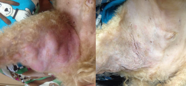
Mast cell tumor (MCT) is the most common skin cancer found in dogs. MCTs originate from the overgrowth of mast cells, one of the normal immune system cells in the body. Normally, mast cells are triggered to aid in the body’s allergic and inflammatory responses. In a cancerous process, however, the mast cells replicate at a much higher rate and become very numerous within the tumor. This may lead to skin irritation and itchiness around the mass itself, as well as gastrointestinal upset due to stomach ulcer formation. Similar to other tumor types, MCTs can metastasize (spread) to other skin sites, lymph nodes, and distant organs.
If an itchy, ulcerated skin mass is noted on your dog what should you do?
The first step to diagnosing a MCT is to have an evaluation performed by your veterinarian. Your veterinarian will decide which method of sampling for the mass is warranted. MCTs can be diagnosed via fine needle aspirate (FNA), or surgical biopsy with analysis of the mass by a pathologist. An FNA of the mass will be able to determine if the mass is indeed a MCT, while a biopsy and analysis of the mass by a pathologist allows the mass to be graded. Grading of the tumor is vital to determine the next treatment steps.
MCTs are graded to correspond with their aggressiveness. A low grade, or grade I, MCT is more likely to remain local and not spread to the lymph nodes or distant organs. A high grade, or grade III, MCT is more likely to behave aggressively and spread to the lymph nodes and distant organs. If a MCT is determined to fall somewhere in between a high and low grade (intermediate or grade II), other factors must be considered to determine the potential aggressiveness of the tumor and appropriate treatment recommendations.
Prior to surgery, various staging options will be discussed and recommended. Typically, an FNA of the lymph node nearest to the site of the mass should be performed to determine if the MCT has spread. An abdominal ultrasound may also be recommended in certain patients to evaluate for MCT spread to distant organs, such as the spleen. While MCTs do not generally metastasize to the lungs, chest radiographs (x-rays) are always a good idea prior to surgery and anesthesia to evaluate the heart and lungs. Blood work is useful to determine any underlying conditions and to ensure your pet is healthy enough for surgery.
Surgical removal of the MCT, when feasible, is recommended as the first step in treatment. In a low grade MCT, complete surgical removal of the tumor (confirmed by the biopsy report) is generally considered curative and your pet will only need to be rechecked periodically with physical exams in the future. In a high grade tumor that is completely surgically excised, chemotherapy is generally recommended. The first line therapy used at Metropolitan Veterinary Associates involves using an injectable chemotherapy protocol consisting of a drug called vinblastine along with an oral steroid (prednisone). A protocol of eight total treatments is used in patients that have already had surgery to remove their mast cell disease. While chemotherapy does have potential side effects, it is generally extremely well tolerated in dogs with a very low risk for stomach upset. Definitive (curative intent) radiation therapy may also be an option for tumors that have been incompletely removed to try to prevent future regrowth.
If a MCT is not able to be surgically excised, there are still options. Chemotherapy may be used to shrink or maintain the size of the mass. An alternative option is an oral medication called Palladia. This drug was developed by Pfizer to treat high grade MCTs. Another option is a type of radiation therapy called palliative radiation. A consultation with an oncologist can help you to decide which options would be best for you and your individual pet.
While cancer can seem a frightening prospect for any pet owner, many options exist for tumors like MCTs to keep your dog happy and comfortable. If you are concerned about a new lump or bump on your pet, have it checked out by your primary veterinarian. If necessary, a referral to an oncologist can be helpful to confirm a diagnosis, review available treatment options, and develop a plan that works for you and your pet.
Before and After Treatment

