A Pet Owner’s Guide to CCL Tears in Dogs
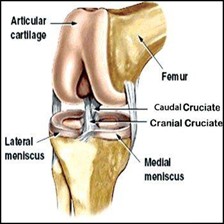
Dogs and humans share a very important but easily damaged joint. In people it is called the knee. In dogs it is called the stifle. The cranial cruciate ligament, or CCL, in the dog is the equivalent of the anterior cruciate ligament, or ACL, in people.
- Cranial cruciate ligament injury is the most common cause of rear limb lameness.
- Dogs can tear or rupture this ligament when the joint is rotated or overextended.
- Obesity, biomechanical problems, or repeated minor stresses can also take a toll on the ligament, causing harmful
If you’re worried your dog might have a CCL tear, you are in the right place. This guide will walk you through the common signs, explain the best treatment options, and set clear expectations for surgery and recovery.
How to Recognize a CCL Tear: Common Signs
- Limping: This is the most common sign of a CCL injury
- Abnormal sitting posture: Your dog may sit with the injured leg held out to the side
- Trouble Getting Up: Difficulty rising from a resting position
- Stiffness: The gait may appear stiff, especially after rest
- Exercise intolerance
- Muscle Atrophy: The muscles in the injured hindlimb may weaken and shrink
- Stifle swelling and pain
What Causes a CCL Tear?
Large active dogs and those that are overweight are more prone to CCL injury. Cats and small dogs can also rupture the CCL, but the incidence is lower and more likely to occur later in life.
Injury to the CCL can be complete or partial “rupture.” If left untreated, the ruptured ligament and resultant joint instability lead to joint swelling, pain and arthritis. When this occurs in one joint, it then places additional stress on the opposite hindlimb as the dog compensates for the resulting pain. This often causes degenerative changes in the opposite stifle (knee) as well. A dog with CCL rupture in one stifle has a higher chance of developing the same problem in the opposite stifle within 1-2 years (approximately 50%).
Treatment Options for a CCL Tear in Dogs
Treatment for this painful condition requires surgical intervention in the vast majority of cases. The goal of treatment is to stabilize the joint to allow normal joint movement, thereby alleviating the dog’s pain and allowing for normal activity and a happy, healthy quality of life.
Non-Surgical (Conservative) Management
Small dogs (under 10 kg or 22 lbs) and can very occasionally be managed with conservative treatment consisting of:
- Exercise moderation
- Non-steroidal anti-inflammatory pain relievers like Deramaxx, Metacam, Previcox, and Rimadyl. In general, these medications provide pain relief and are anti-inflammatory. All drugs in this group have the potential for causing gastro-intestinal irritation, so if your pet has any vomiting or diarrhea whilst on this medication you should stop giving it and contact your veterinarian.
- Joint supplements such as Glucosamine and chondroitin sulfate
- May also include a custom stifle brace
However, it is important to note that even with this approach, intermittent discomfort continues along with the progression of degenerative joint disease (arthritis).
Surgical Treatment: Comparing Your Options
At Metropolitan Veterinary Urgent Care and Specialty, we offer multiple surgical options:
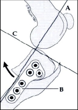
- Tibial Plateau Leveling Osteotomy (TPLO) with arthroscopic joint evaluation:
Many animals with cruciate ligament injury have a sloped top to their tibia (shin bone). This slope puts excess stress on the cruciate ligament and contributes to it rupturing. The TPLO surgery works by altering the biomechanics of the joint and eliminating the need for a CCL. During surgery, the top of the tibia is cut and rotated to a new position to level the top of the tibia, creating a level weight bearing platform. It is held in the new position using a bone plate and screws. This technique is used most frequently in medium to giant breed dogs, obese dogs, performance dogs and dogs with cruciate injury in both legs.In our experience, dogs tend to bear weight on their operated leg a little quicker after the TPLO surgery than they do with the extracapsular technique. This is of benefit when they have cruciate ligament injury in their other knee. - Extracapsular stabilization technique:
This technique seeks to replace the function of the CCL with a prosthetic ligament outside of the joint oriented in the same direction as the previous CCL. In addition, the tough tissue outside the joint (fascia) is tightened to provide additional stability to the joint. This prosthetic suture is a temporary stabilization, since over time the material stretches and can ultimately break. The success of this technique relies on the development of fibrous tissue around the joint that takes over the function of the prosthetic and stabilizes the joint. It is more commonly used in smaller dogs and cats. This method is commonly referred to as a “lateral suture.”This is a very strong fixation and less likely to have implant failure than the extracapsular technique, so it is of benefit in very large dogs or dogs that are overweight. However, failure of the implants (breakage of the plate or screws or pull out of the screws from the bone) is possible and is more difficult to deal with than complications associated with the extracapsular technique.
Why we don’t offer TTA Surgery: We do not perform the Tibial Tuberosity Advancement (TTA) technique due to published data showing inferiority to the TPLO procedure.
With either the extracapsular stabilization or the TPLO technique, the first step of the surgery is the same — visually exploring the stifle joint. This visualization can be performed with an open approach to the joint or an arthroscopic approach, using a small camera to visualize and operate within the joint. After visualizing the cranial cruciate ligament, damaged portions will be removed as it releases inflammatory mediators into the joint that cause ongoing pain, lameness, and progression of arthritis.
We also evaluate the meniscal cartilages inside the joint. These cartilages act as shock absorbers and can become damaged when the cruciate ligament is damaged. If there is meniscal injury, the damaged portion of the meniscus is removed. In some cases, the meniscal tear can be repaired. If the meniscal cartilages are not damaged, they are left in place since they provide an important role in the joint – however, dogs do have a slight risk of developing an isolated meniscal injury in the future, requiring re-exploration of the joint.
Success Rates Potential Complications
The success rate for both TPLO and Extracapsular Stabilization is high, currently between 85-95%. Your pet should be able to return back to normal, or near normal, activity over a 2-4 month period. There are a small percentage of dogs and cats that do not do well following cruciate ligament injury, no matter how they are treated.
However, as with any surgery, there are risks involved. The most common complications include:
- Anesthesia: There is always a risk with anesthesia although it is rare that we have significant problems. All animals at MVUCS are anesthetized with a custom tailored protocol and are continuously monitored by a veterinary nurse throughout the procedure. They are placed on intravenous fluids and their heart rates, respiratory rates and blood pressure are constantly monitored. They are also placed on a circulating warm water blanket to help maintain their body temperature under anesthesia.
- Infection: There is always the potential for infection with any surgery. Our infection rate is extremely low. In most cases, infection occurs when the animal licks or chews at the surgical incision postoperatively. To minimize this, we send your pet home with a protective collar that they should wear at all times to prevent licking or chewing at the incision. With either surgery- if an infection develops it can delay healing and necessitate the removal of the implants (the stabilizing ligament or the bone plate and screws), which is additional anesthesia and surgery for your pet and additional cost.
- Implant failure: Premature breakdown of the stabilizing extracapsular prosthetic ligament can occur and is more likely in very large, active dogs or obese dogs. It is also more likely if dogs do not have their activity restricted as directed postoperatively. If the material breaks prematurely it can lengthen the recovery process and in some cases necessitate further surgery. The bone plate and screws used in the TPLO are very strong but by cutting the bone – this is essentially the same as having a fractured bone that requires time to heal. Initially the strength of the repair is provided by the plate and screws alone. Over the next few weeks, as the bone begins to heal, the bone starts providing additional strength to the repair. It is rare that we have implant failure with this technique. If the plate or screws break, the plate pulls off the bone, or the bone breaks around the screws, further surgery is required and can be very difficult to perform.
- Isolated meniscal injury: As previously stated, dogs do have a slight risk of developing an isolated meniscal injury in the future, requiring re-exploration of the joint. This can occur with either technique, and it is not possible to predict which dogs will be affected.
- Tibial Tuberosity fracture (fracture through the top, front part of the tibia or shin bone): This is a potential complication of the TPLO procedure. It occurs infrequently but depending on how long it occurs after the surgery and the displacement of the piece of bone that breaks, it may require placement of metal pins to repair the damaged piece of bone.
- Patellar Tendon Inflammation: This is an uncommon complication following the TPLO procedure and can prolong the recovery period. It does not require surgery but can necessitate a prolonged period of activity restriction.
- Persistent Lameness secondary to osteoarthritis: This affects between 10-15% of dogs. This is a potential complication with either surgery, and there is no way to predict which dogs will have significant problems with arthritic pain. It requires life-long activity moderation (less free running and jumping, more slow leash walks and swimming) and the use of non-steroidal anti-inflammatory drugs long term (drugs such as Rimadyl, Deramaxx, Metacam and Previcox).
Post-Operative Care: Your Role in a Successful Recovery
Specific instructions will be given at the time your pet is discharged from the hospital including what medications to give. In general, animals have their incisions checked 12-14 days after surgery, either in person or virtually.
Following surgery your pet will require strict activity restriction. Patient activity is generally restricted for at least 2.5-3 months.
Weeks 1 and 2 post operatively: For the first 12-14 days your dog should be confined to a crate or small room with minimal furniture. Steps should be kept to a minimum. They should go outside on a short leash (not at the end of an extendible leash) to go to the bathroom only – no free running, jumping or playing is allowed. This can result in failure of the implants and may result in the need for more surgery.
If your dog will tolerate it you can apply an ice pack to the incision up to 4 times daily for 3-5 minutes for the first 3-5 days. Do not try to force this if your dog will not let you.
Once the incision has healed, you can start the following walking program at home – all other restrictions still apply and your dog should still not be allowed to roam free in the house:
Week 3 post operatively: Slow, on a short leash walks for 5 minutes up to 3-4 times daily. You can also start making your dog walk in some small circles and figures of 8 with the operated leg on the inside of the circle/8.
Week 4 postoperatively: Slow, on a short leash, walks for 10 minutes up to 3-4 times daily.
Week 5 postoperatively: Slow, on a short leash walks for 15 minutes up to 3-4 times daily.
Week 6 postoperatively: Slow, on a short leash walks for 20 minutes up to 3-4 times daily.
For dogs that have had a TPLO, weeks 7 and 8 should be the same as week 6: Slow, on a short leash walks for 25 minutes up to 3-4 times daily.
If your dog is hard to control on the leash and is liable to injure themselves their activity should be completely restricted to leash walks to the bathroom only for the first 6 weeks.
Following extracapsular stabilization, dogs are then seen for a recheck at 6 weeks; further activity instructions will be given at this visit.
Dogs that have TPLO performed come back for follow-up X-rays at 8 weeks to make sure that the cut in the bone is healing. There is an additional charge for these X-rays and sedation. If the cut in the bone has not healed at 8 weeks, further X-rays will be taken at 12 weeks postoperatively. Dogs that had extracapsular stabilization do not require follow-up X-rays in most cases.
All pets with stifle problems should maintain an ideal body weight and you may need to decrease the amount you are feeding during the postoperative recovery period.
The amount of arthritis that an animal develops after cruciate ligament injury is variable and there is no way to predict how it will affect your pet. It is important that once they have healed from surgery that they have regular exercise, maintain an ideal body weight and, if possible, stay on a Glucosamine/Chondroitin joint supplement for life.
In addition, some animals may require the tactical use of non-steroidal anti-inflammatory drugs such as Rimadyl, Deramaxx, Previcox or Metacam. These can be obtained from your regular veterinarian and be used on an “as needed” basis. Arthritis tends to cause more discomfort after periods of sudden very heavy exercise (free running, jumping, ball chasing) and when the weather is colder and damper.
Is your Dog showing signs of a CCL tear or injury? Contact Metropolitan Veterinary Urgent Care and Specialty today to schedule a consultation with our surgical team.